News, Research and other Resources:

+ Frequently asked questions about Mandibular Advancement Therapy.

Dr Keith Burgess (Sleep Physician) has been prescribing Mandibular Advancement Devices (MAD’s) for 25 years and has taken the time to answer some of the common questions asked by patients considering the use of a MAD for the treatment of their Obstructive Sleep Apnoea and/or snoring.

1. What severity of obstructive sleep apnoea (OSA) is suited to having a two piece adjustable mandibular advancement device (MAD)?

The best suited are those patients with “simple snoring” or mild to moderate severity OSA.

2. What other factors do you consider in recommending MAD? (E.g: weight, tongue size snoring etc…)

There are data that suggest that obese patients do not do as well as the non obese.
But, that is less of an issue than which treatment will the patient accept and use?
There is no point pushing CPAP therapy if the patient will not accept it. Adequate dentition is essential. The patient must have at least 8 strong teeth top and bottom to use a MAD, and the more the better.

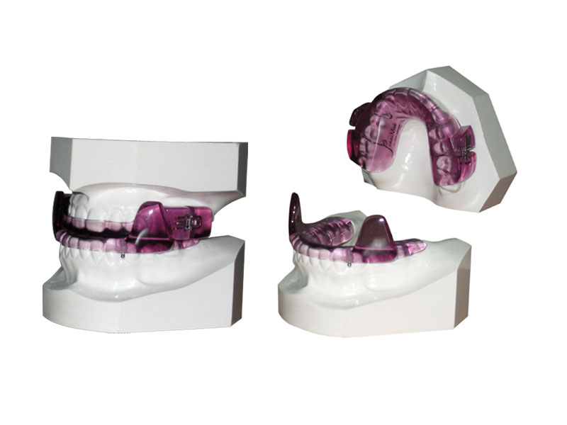
3. What is the process for having a MAD fitted?

Referral to an experienced dentist.
The dentist takes an impression or a computer scan of the patient’s teeth.
The mouldings or computer file are then sent to a lab who manufactures the device.
The patient returns to the dentist, usually three weeks after the assessment, to have a test fit of the MAD. Any discomfort issues are dealt with, and often the device is advanced a little.
The patient then uses it for two weeks without adjustment.
They then return to the dentist for review and another advancement.
The dentist then shows the patient how to advance it and sets up a schedule, or books appointments for further advancements.
Once the snoring stops, or is much reduced, or the patient reaches 2mm in front of “edge to edge” the advancing stops.
The patient is then reviewed by the sleep physician and a follow up sleep study arranged.

4. Is it important to be re-tested once the device is fully adjusted?

Yes. Except for those patients who only had snoring without OSA, who do not need another study. For all those with OSA they should be restudied to make sure that the OSA is being controlled at that degree of advancement. Often a further adjustment is required.

5. What is the rough cost of a device?

The cost depends on the dentist. To my knowledge the fee ranges between $1500 and $3000 which includes the device itself and the dentists' time and expertise.*
*Note these costs are estimations and you will need to discuss your expected costs with your dentist

6. How long do they last?

If the patient does not tread on them nor their dog eat them (common problems apparently) they should last 5 years.

7. Are there any side effects and/or contraindications to MAD treatment?

There are few contraindications: a strong gag reflex and inadequate dentition are the main ones. Temporo-mandibular joint problems are NOT a contra indication.
Most patients will have some degree of bite change due to the device. Most patients do not mind and very few have to stop using it because of marked bite change. Unfortunately, it is not possible to predict who will suffer from this and who will not. In my experience it does not appear to be related to the degree of advancement.

Dr Burgess has no financial interest in any company that manufactures MAD’s. He is the sole Medical Director of Peninsula Sleep Clinic.


+ Influence of Cerebral Blood Flow on Central Sleep Apnea at High Altitude.
Burgess KR, Lucas SJ, Shepherd K, Dawson A, Swart M, Thomas KN, Lucas RA, Donnelly PJ, Peebles KC,Basnyat R, Ainslie PN Peninsula Sleep Laboratory, Sydney, New South Wales, Australia. Sleep [2014, 37(10)]

Abstract

Study Objectives:
To further our understanding of central sleep apnea (CSA) at high altitude during acclimatization, we tested the hypothesis that pharmacologically altering cerebral blood flow (CBF) would alter the severity of CSA at high altitude.

Design:
The study was a randomized, placebo-controlled single-blind study.

Setting:
A field study at 5,050 m in Nepal.

Patients or Participants:
We studied 12 normal volunteers.

Interventions:
Between days 5 to10 at high altitude, CBF velocity (CBFv) was increased by intravenous (IV) acetazolamide (10 mg/kg) and reduced by oral indomethacin (100 mg).

Measurements and Results:
Arterial blood gases, hypoxic and hypercapnic ventilatory responses, and CBFv and its reactivity to carbon dioxide were measured awake. Overnight polysomnography was performed. The central apnea-hypopnea index was elevated following administration of indomethacin (89.2 ± 43.7 to 112.5 ± 32.9 events/h; mean ± standard deviation; P < 0.05) and was reduced following IV acetazolamide (89.2 ± 43.7 to 47.1 ± 48.1 events/h; P < 0.001). Intravenous acetazolamide elevated CBFv at high altitude by 28% (95% confidence interval [CI]: 22-34%) but did not affect ventilatory responses. The elevation in CBFv was partly mediated via a selective rise in partial pressure of arterial carbon dioxide (PaCO2) (28 ± 4 to 31 ± 3 mm Hg) and an associated fall in pH (P < 0.01). Oral indomethacin reduced CBFv by 23% (95% CI: 16-30%), blunted CBFv reactivity, and increased the hypercapnic ventilatory response by 66% (95% CI: 30-102%) but had no effect on PaCO2 or pH.

Conclusions:
Our findings indicate an important role for cerebral blood flow regulation in the pathophysiology of central sleep apnea at high altitude.

Access the full article here.


+ Targeted Case Finding for OSA within the Primary Care Setting.
Journal of Clinical Sleep Medicine.
Keith R. Burgess, Ph.D.,1 Adrian Havryk, Ph.D.,1 Stephen Newton, M.B.A.,2 Willis H. Tsai, M.D., F.A.A.S.M.,3 andWilliam A. Whitelaw, M.D., Ph.D.4
1 Peninsula Respiratory Group, Frenchs Forest, NSW, Australia 2 Healthy Sleep Solutions Pty Ltd, Sydney, NSW, Australia 3 Department of Community Health Sciences, University of Calgary, Calgary, Alberta, Canada 4 Department of Medicine, University of Calgary, Calgary, Alberta, Canada


Abstract

Study Objectives:
The aim was to determine the feasibility of using an unattended 2-channel device to screen for obstructive sleep apnea in a population of high-risk patients using a targeted, case-finding strategy. The case finding was based on the presence of risk factors not symptoms in the studied population.

Methods:
The study took place from June 2007 to May 2008 in rural and metropolitan Queensland and New South Wales. Family doctors were asked to identify patients with any of the following: BMI > 30, type 2 diabetes, treated hypertension, ischemic heart disease. Participants applied the ApneaLink+O2 at home for a single night. The device recorded nasal flow and pulse oximetry. Data were analyzed by proprietary software, then checked and reported by either of two sleep physicians.

Results:
1,157 patients were recruited; mean age 53 +/- 14.6, M/F% = 62/38, mean BMI = 31.8, obesity = 35%, diabetes = 16%, hypertension = 39%, IHD = 5%, Mean Epworth Sleepiness Scale score (ESS) = 8.3. The prevalence of unrecognized OSA was very high: 71% had an AHI > 5/h, 33% had an AHI > 15/h, and 16% had an AHI > 30/h. The ApneaLink+O2 device yielded technically adequate studies in 93% of cases.

Conclusions:
The study shows that a "real world" simple low cost case finding and management program, based on unattended home monitoring for OSA, can work well in a population with risk factors and comorbidities associated with OSA, independent of the presence of symptoms. The prevalence of unrecognized OSA was very high.

Access the full article here.


+ Breathing and sleep at high altitude.
Journal of Respiratory Physiology and Neurobiology.
Ainslie PN1, Lucas SJ2, Burgess KR 3,4.
1 Centre of Heart, Lung and Vascular Health, School of Health and Exercise Sciences, University of British Columbia, Okanagan Campus, British Columbia, Canada. 2 School of Sport, Exercise and Rehabilitation Sciences, College of Life and Environmental Sciences, University of Birmingham, Edgbaston, Birmingham, UK. 3 Peninsula Sleep Clinic, Sydney, New South Wales, Australia. 4 Department of Medicine, University of Sydney, Sydney, New South Wales, Australia.


Highlights

• Ventilatory acclimatization to altitude involves cellular and neurochemical re-organization in the peripheral chemoreceptors and CNS.
• Sleep at high altitude is disturbed by various factors, but principally by periodic breathing (PB).
• The extent of PB during sleep at altitude intensifies with duration and severity of exposure and is explained in part by elevations in loop gain.
• Because PB may elevate rather than reduce mean SaO2 during sleep this may represent an adaptive rather than maladaptive response.
• Although new mechanical and pharmacological means are emerging, oral acetazolamide remains the most effective and practical means to reduce PB.

Abstract:
We provide an updated review on the current understanding of breathing and sleep at high altitude in humans. We conclude that: (1) progressive changes in pH initiated by the respiratory alkalosis do not underlie early (<48 h) ventilatory acclimatization to hypoxia (VAH) because this still proceeds in the absence of such alkalosis; (2) for VAH of longer duration (>48 h), complex cellular and neurochemical re-organization occurs both in the peripheral chemoreceptors as well as within the central nervous system. The latter is likely influenced by central acid-base changes secondary to the extent of the initial respiratory responses to initial exposure to high altitude; (3) sleep at high altitude is disturbed by various factors, but principally by periodic breathing; (4) the extent of periodic breathing during sleep at altitude intensifies with duration and severity of exposure; (5) complex interactions between hypoxic-induced enhancement in peripheral and central chemoreflexes and cerebral blood flow – leading to higher loop gain and breathing instability – underpin this development of periodic breathing during sleep; (6) because periodic breathing may elevate rather than reduce mean SaO2 during sleep, this may represent an adaptive rather than maladaptive response; (7) although oral acetazolamide is an effective means to reduce periodic breathing by 50–80%, recent studies using positive airway pressure devices to increase dead space, hyponotics and theophylline are emerging but appear less practical and effective compared to acetazolamide. Finally, we suggest avenues for future research, and discuss implications for understanding sleep pathology.

Access the full article here.


+ Worsening of central sleep apnoea at high altitude - a role for cerebrovascular function.
KEITH BURGESS, 1. 2. SAMUEL J E LUCAS, 3. KELLY SHEPHARD, 1. ANDREW DAWSON, 1. MARIANNE SWART, 1. KATE N THOMAS, 3. REBEKAH A I LUCAS, 3. JOSEPH DONNELLY, 3. KAREN C PEEBLES, 3. RISHI BASNYAT, 4, 5. and PHILIP N AINSLIE, 6.
1. Peninsula Sleep Laboratory, Sydney, New South Wales, Australia; 2. Department of Medicine, University of Sydney, Sydney, New South Wales, Australia; 3. University of Otago, Dunedin, New Zealand; 4. Nepal International Clinic, Kathmandu, Nepal; 5. Banner Good Samaritan Hospital Medical Centre, Phoenix Arizona; and 6. Centre for Heart, Lung and Vascular Health, School of Health and Exercise Sciences, University of British Columbia, Okanagen Campus, Canada.


Abstract

Background and Methods:
Although periodic breathing during sleep at high altitude occurs almost universally,the likely mechanisms and independent effects of altitude and acclimatization have not been clearly reported. Data from 2005 demonstrated a significant relationship between decline in cerebral blood flow (CBF) at sleep onset and subsequent severity of central sleep apnoea that night. We suspected that CBF would decline during partial acclimatization. We hypothesized therefore that reductions in CBF and its reactivity would worsen periodic breathing during sleep following partial acclimatization.

Methods:
Repeated measures of awake ventilatory and CBF responsiveness, arterial blood gases during wakefulness.and overnight polysomnography at sea level, upon arrival(days 2–4), and following partial acclimatization (days 12–15) to5,050 m were made on 12 subjects.

Results:
The apnea-hypopnea index (AHI) increased from to 77  49 on days 2–4 to 116  21 on days 12–15(P  0.01). The AHI upon initial arrival was associated with marked elevations in CBF (28%, 68  11 to 87  17 cm/s; P  0.05) and its reactivity to changes in PaCO2 [90%, 2.0  0.6 to 3.8  1.5cm·s1·mm Hg1 hypercapnia and 1.9  0.4 to 4.1  0.9cm·s1·mm Hg1 for hypocapnia (P  0.05)]. Over 10 days, the increases resolved and AHI worsened. During sleep at high altitude large oscillations in mean CBF velocity (CBFv) occurred, which were 35% higher initially (peak CBFv  96 cm/s vs. peak CBFv  71cm/s) than at days 12–15.

Conclusions:
Our novel findings suggest that elevations in CBF and its reactivity to CO2 upon initial ascent to high altitude may provide a protective effect on the development of periodic breathing during sleep (likely via moderating changes in central PCO2).

Access the full article here.


+ The effect of partial acclimatization to high altitude on loop gain and central sleep apnoea severity.
GARETH ANDREWS, 1, 2. PHILIP N. AINSLIE, 3. KELLY SHEPHERD, 2. ANDREW DAWSON, 2. MARIANNE SWART, 2. SAMUEL LUCAS, 3. AND KEITH R. BURGESS, 1.
1. Department of Medicine, University of Sydney, 2. Peninsula Respiratory Group, Sydney, New South Wales, Australia, and 3. Department of Physiology, University of Otago, Dunedin, New Zealand


Abstract

Background and Objective:
Loop gain is an engineering term that predicts the stability of a feedback control system, such as the control of breathing. Based on earlier studies at lower altitudes, it was hypothesized that acclimatization to high altitude would lead to a reduction in loop gain and thus central sleep apnoea (CSA) severity.

Methods:
This study used exposure to very high altitude to induce CSA in healthy subjects to investigate the effect of partial acclimatization on loop gain and CSA severity. Measurements were made on 12 subjects (age 30 ± 10 years, body mass index 22.8 ± 1.9, eight males, four females) at an altitude of 5050 m over a 2-week period upon initial arrival (days 2–4) and following partial acclimatization (days 12–14). Sleep was studied by full polysomnography, and resting arterial blood gases were measured. Loop gain was measured by the ‘duty cycle’ method (duration of hyperpnoea/cycle length).

Results:
Partial acclimatization to high-altitude exposure was associated with both an increase in loop gain (duty cycle fell from 0.60 ± 0.05 to 0.55 ± 0.06 (P = 0.03)) and severity of CSA (apnoea-hypopnoea index increased from 76.8 ± 48.8 to 115.9 ± 20.2 (P = 0.01)), while partial arterial carbon dioxide concentration fell from 29 ± 3 to 26 ± 2 (P = 0.01).

Conclusions:
Contrary to the results at lower altitudes, at high-altitude loop gain and severity of CSA increased.

Access the full article here.


+ Peninsula Sleep Laboratory CSL Biotherapies education meeting 2012.
Peninsula Sleep Laboratory (PSL) in conjunction with CSL Biotherapies sponsored PSL's annual educational meeting, held at the Novotel Manly beachfront on Tuesday 14th of August. The night had a great atmosphere with 45 local general practitioners, cardiologists, ear nose and throat and respiratory and sleep specialists turning out. World class speakers Professor Ian Willcox and A/Prof Stuart Mackay entertained with cutting edge medical sleep research and practical approaches to patient treatment.

Professor Wilcox presented Sleep Apnoea as an adverse risk factor for cardiac arrhythmias and their treatment. A/Prof Stuart MacKay presented surgical solutions for adult snoring and OSA.

Peninsula Sleep Laboratory holds this event annually for local doctors to attend and grow their understanding in the field of sleep medicine. For any interest please contact the Laboratory directly. The next event will be around July 2013.


+ Peninsula Sleep Laboratory in the Himalaya 2012!


Peninsula Sleep Laboratory has returned from its international six weeks research expedition in the Himalaya. After a 10 day hike up to the EV-K2-CNR Italian Research Pyramid (http://www.evk2cnr.org) at 5050 meters in the Kumbu Valley. Several days to acclimatize were required before embarking on 18 nights in a row of sleep studies. Twenty three researchers conducted almost as many projects over the 21 days at the Pyramid to expand our understanding of how the human body works, particularly in a physiological sense, so that new treatments for sea level and high altitude diseases may be developed.

Professor Keith Burgess was the Principle investigator on two different sleep projects in conjunction with Professor Phil Ainslie (Canada), Dr Sam Lucas (New Zealand), Dr Jim Cotter (New Zealand), Dr David McLeod (USA) and Dr Aparna Basnet (Nepal). This research included a follow up study of sleep during alteration of cerebral blood flow which they first attempted in 2008 at the Pyramid. In addition to this a new study of central sleep apnea during different pH conditions was conducted.

High altitude induces central sleep apnea in most subjects which provides a stable model with which to work for these projects. The insights that we gain from this model may be able to be applied to sick patients at sea level who also have central sleep apnea. In addition it stresses the normal human physiology in ways that do not occur at sea level. Studying normal subjects at high altitude may provide new understandings of how the normal human body works.

A mobile sleep lab was setup with the help of Compumedics (www.compumedics.com) who provided blue tooth portable Polysomnography equipment for live acquisition. Four sleep studies per night were conducted in camp style beds with trans-cranial cerebral blood flow monitoring and arterial blood samples were taken during sleep. Ventilatory responses to hypoxia and hypercapnia were studied during the daytime with cardiac and vascular function analysis simultaneously.

Many months of analysis will now begin unraveling the data collected in order to gain a better understanding of mechanisms behind central sleep apnoea. Watch this space for more information.


+ Compumedics Australian News 2012


The Peninsula Sleep Laboratory in New South Wales has been involved in sleep research at high altitude levels since 1997. The Medical Director Dr. Keith Burgess and the Laboratory Manager Katie, will be returning to the Nepal Himalaya mountains in April and May of this year, to conduct further research into sleep at high altitude.

Part of an international expedition of over 20 members from Canada, Australia and New Zealand, they will be using a joint Italian and Nepali Research Station at 5,050 metres called "The Pyramid". The Station was established in 1994 to study climatic change and earth movement, but has been used by numerous investigators since for physiology experiments as well.

The international expedition will study a number of physiological questions related to high altitude exposure and physiological adaptation, including the development of central sleep apnoea, which is an almost universal occurrence at that altitude. Arterial blood gas changes, ventilatory responses, cerebral blood flow changes and the physiology of sleep will be measured. During earlier experiments in 2008, the same investigators discovered novel findings associated with acclimatisation. This time they will be trialling potential new interventions in the treatment of central sleep apnoea, that might have useful ramifications at sea level.

To study sleep, the team will be using four Compumedics Somté PSG battery powered polysomnography systems - these same systems worked extremely well during the study conducted in 2008. The set up this year will be slightly different as the team need to be able to record to SD cards, but monitor live acquisition on two laptops overnight. The study will be done with the aim of watching this live to see effects present whilst they administer drugs at different points during the night.

The investigating team are grateful to Compumedics for their support of the expedition, through the provision of the Somté PSG systems.alia


+ An Objective Assessment Of The Effectiveness Of Mandibular Advancement Devices In Home Use.
Keith Burgess (1,2), Andrew Burgess (2,3), Hedi Lamy (2), (1). Department of Medicine, University of Sydney. Sydney. NSW. 2050, (2). Peninsula Sleep Laboratory. Sydney. NSW. 2086, (3). Faculty of Medcine, Notre Dame University. Sydney. NSW. 2010, Australia

Abstract

Background:
Mandibular Advancement Devices (MADs) are now used more frequently to treat Obstructive Sleep Apnoea (OSA). Their effectiveness in practice is uncertain. Currently they are often thought of as providing treatment success in only 50% of patients. We suspected that using a second polysomnogram (PSG) to guide final adjustment would improve effectiveness.

Aim:
To objectively assess in the home the effectiveness of MADs in moderate severity OSA using our paradigm. Methods: We invited 200 subjects treated by the same algorithm to be restudied in their home environment. [Paradigm = Initial PSG, MAD advanced till “snoring controlled”, repeat PSG, further adjustment based on 2nd PSG]. 50 subjects (age 63±10, M:F 33:17) were studied with the MAD insitu by unattended PSG in their homes. (Somte PSG. Compumedics. Melbourne). All studies were scored by a certified technician not involved in the study, using R & K rules and “Chicago criteria”. (Snoring loudness was scored: 0=nil, 1=mild, 2=moderate, 3=loud, 4=very loud). The home studies with MAD in situ were compared to the original PSGs for sleep related variables and potential confounders. A variety of devices were used, though 65% were Somnodent. Treatment success was defined as normalisation of AHI or more than 50% reduction in AHI.

Results:
Total sleep time increased from 331±73 mins to 381±56 (P<001). Despite an increase in REM sleep from 15.8 ± 5.4% to 17.5 ± 4.9% (P=0.02), arousal index fell from 28 ± 13/hr sleep to 17 ± 8/hr at home (P<0.001). AHI fell from 21.8 ± 14/hr to 9.5 ± 9.8/hr (P<0.001), Desaturation below 90% fell from 4.7±12.4% sleep time to 2.3±4.9% (P=0.1). Snoring Score was reduced from 2.5 ± 0.7 to 1.7 ± 0.8 (P<0.001), BMI was unchanged at 28.6 ± 4.2. Supine sleep % was unchanged @ 38±26.

Conclusions:
In the home environment MADs reduced RDI by 56%, & desaturation below 90% by 51%. Treatment success occurred in 65% initially then in 70% subjects after final adjustment. There were no identified confounders. Conflict of interest: Yes


+ Peninsula Sleep Laboratory Educational Evening "Sleep. Who needs it?" – March 2nd 2010
Clinical Professor Keith Burgess MB BS, M.Sc, Ph.D, FRACP, FRCP, FACP (Respiratory and Sleep Disorders Physician) talked about "Manipulating Sleep at High Altitude" and "New Data on Dental Devices in OSA". Dr Dianne Richards B Soc. Sc (Sleep Psychologist) spoke about "Non Pharmacological Treatment for Insomnia". The evening attracted 40 General Practitioners, Dentists and Specialists and was a great success.


+ Transformation of Obstructive Sleep Apnoea at Sea Level to Central Sleep Apnoea at High Altitude; Influence of Cerebral Blood Flow
Under special circumstances Obstructive Sleep Apnoea (OSA) and Central Sleep Apnoea (CSA) can occur in the same patient at different times; the transformation of OSA at sea level to CSA at high altitude and simulated high altitude have been reported (1). Those reports lacked measures of ventilatory response or cerebral blood flow that might help explain the underlying physiological mechanisms. Here, we report data from one otherwise healthy subject (54 years) who participated in experiments investigating the effects of pharmacological-induced alterations in cerebral blood flow velocity (CBFv) during sleep monitored with full polysomnography. At sea-level he had mild OSA (AHI = 14/hr) which was completely resolved at high altitude (5,050m) and replaced with severe CSA (AHI = 108/hr). During wakefulness, whilst his resting CBFv was unaltered at high altitude from that at sea-level, his cerebrovascular response to CO2 was reduced by 38 % and the ventilatory response to hypercapnia was elevated (0.1 to 1.0 l/min/mmHg); PaCO2 fell from 40 to 25 mmHg following ascent to altitude. Since reductions in CBF-CO2 sensitivity are important determinates of eupnoeic ventilation, hypercapnic ventilatory sensitivity and breathing stability, these factors may partly explain the exacerbation of CSA. Although the mechanisms by which OSA is replaced with CSA at altitude are unclear, hypoxic-induced alterations in chemoreflex stability and upper airway muscle activity are likely to be critical factors.

1. Burgess et al Respirology 2004

This study was supported by the Otago Medical Research Foundation, Peninsula Health Care p/l, Air Liquide p/l and the Italian National Research Council who kindly provided use of the EV-K2-CNR research laboratory.


+ Separate Effects Of Acclimatisation And Cerebral Blood Flow On Central Sleep Apnea At High Altitude
Keith R. Burgess, Andrew Dawson, Kelly Shepherd, Marianne Swart, Kate N. Thomas, Jui-Lin Fan, Rebekah A. I. Lucas, Samuel J. E. Lucas, James D. Cotter, Karen C. Peebles, Rishi Basnyat, Philip N. Ainslie. University of Sydney, Sydney, NSW, Australia. Peninsula Sleep Laboratory, NSW, Australia; University of Otago, Dunedin, New Zealand; Nepal International Clinic, Kathmandu, Nepal.

Exposure to high altitude causes a universal increase in central sleep apnea (CSA), mediated by alterations in ventilatory control and possibly in cerebral blood flow (CBF). The extent to which CSA changes over time at high altitude, and the extent to which it can be altered by pharmacologically induced alterations in CBF is unclear.

We hypothesised that partial acclimatisation and pharmacologically induced alteration of CBF would have separate effects on the frequency and duration of central apneas during sleep at high altitude. We studied 12 normal volunteers on four occasions over a three week period at 5050m, at Lobuje in northern Nepal. Measurements included overnight polysomnography with transcranial Doppler measurement of CBF, non invasive hemodynamics and ABG analysis at the Pyramid Research Station. They were studied at the beginning and end of their stay, to control for acclimatisation, and in between the control nights they were studied after pharmacological intervention. All subjects received oral Indomethacin 100mg and iv Acetazolamide (10mg/kg) 2 hours before sleep, in random order with placebo controls, at approximately 4 day intervals. The data from the pharmacological intervention nights were compared to the mean data from the control nights.

After Indomethacin, CBF fell by 22 ± 8% and the apneas lengthened from 13.9 ± 2.2 to 15.4 ± 3.3secs (p<0.01). Central Sleep Apnea Index (CSAI) increased from 96.4 ± 30.3 to 101 ± 28.2 apneas/hr (NS).

After Acetazolamide, CBF increased by 31 ± 6% , which had no effect on apnea duration but the CSAI fell from 96.4 ± 30.3 to 53.7 ± 45.7 apneas/hr (p<0.001).

During partial acclimatisation, CSAI increased from 76.9 ± 48.9 to 115.9 ± 20.2 /hr over the 12 day period (p=0.01), and apnea duration lengthened from 13.1 ± 2.6 to 14.6 ± 2.2 secs (p<0.02). Over the same period PaCO2 declined from 29±3 to 26±2mmHg, and the rise (25±10%) in CBF upon initial exposure (days 1-4) returned to its sea-level values. We propose that the increase in apnea length with Indomethacin was due to reductions in CBF and cerebrovascular reactivity and increase in “loop gain”. However the lengthening of the apneas due to acclimatisation must be due to mechanisms that are independent of CBF.

This study was supported by the Otago Medical Research Foundation, Peninsula Health Care p/l, Air Liquide p/l and the Italian National Research Council who kindly provided use of the EV-K2-CNR research laboratory.


Back to Top